2026 Best Bone Biopsy Procedure Types to Know?
Choosing the right Bone Biopsy Procedure can shape every later treatment decision. It can distinguish infection, benign lesions, primary sarcoma, and metastatic disease. The biopsy is not merely a tissue sample. It is a carefully planned pathway through bone, muscle, imaging, pathology, and surgery.
The American Cancer Society’s Cancer Statistics 2025 estimates thousands of new primary bone and joint cancer cases annually in the United States. SEER data also shows that these tumors remain uncommon, which makes specialist interpretation especially important. Meanwhile, the WHO Classification of Tumours, Soft Tissue and Bone Tumours, 5th Edition, stresses integrated diagnosis using morphology, immunohistochemistry, and molecular findings. Commercial analyses, including Grand View Research’s bone biopsy market reporting, identify growing demand for image-guided and minimally invasive sampling. Yet market growth does not replace clinical judgment.
Orthopedic oncologist William F. Enneking is widely associated with this principle: “The biopsy is the most important surgical procedure in the management of a patient with a musculoskeletal tumor.” That warning still matters. A poorly positioned tract may complicate limb-sparing surgery. A small, crushed specimen may delay diagnosis. An experienced multidisciplinary team usually selects between image-guided core biopsy, fine-needle aspiration, and open biopsy according to lesion location, vascularity, imaging features, and suspected pathology. The choice is not always obvious. Sometimes, the safest-looking option is not the most informative one. This guide examines the 2026 Bone Biopsy Procedure types clinicians and patients should understand, including their practical advantages, limitations, preparation needs, and diagnostic roles.
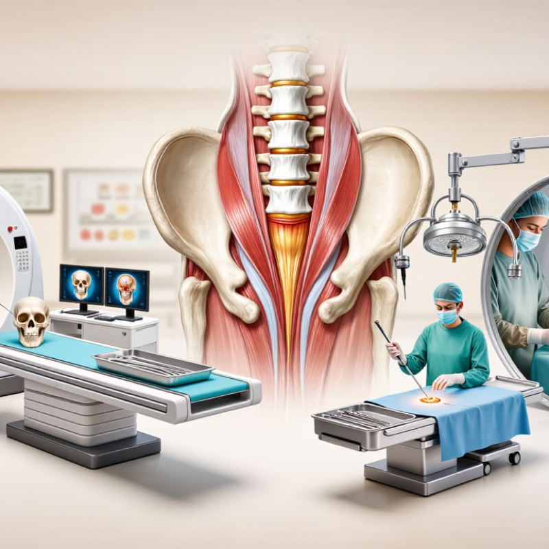
What a Bone Biopsy Is and Why It Is Performed
A bone biopsy removes a small bone or marrow sample for laboratory examination. Doctors may recommend it when imaging shows an unusual lesion, unexplained bone pain, infection, or a possible tumor. The procedure can help distinguish cancer from inflammation, infection, or a noncancerous growth. Imaging alone may suggest a diagnosis, but it cannot always provide a reliable answer.
There are two common approaches. A core needle biopsy uses a hollow needle, often guided by computed tomography, to collect tissue through a small skin opening. An open biopsy requires a surgical incision and may be chosen when the lesion is difficult to reach or a larger sample is needed. During a needle procedure, patients may feel pressure, vibration, or brief discomfort after local anesthetic. Some people still feel anxious. That matters.
Before the biopsy, the medical team reviews scans, medicines, allergies, bleeding risks, and the planned sampling route. The tissue then goes to a pathology laboratory, where specialists examine its cells and structure. Results may take several days, especially when infection tests or additional studies are needed. A biopsy is highly useful, but it is not flawless; a small sample can miss important tissue. Persistent pain, swelling, fever, drainage, or worsening redness after the procedure requires prompt medical advice.
How Doctors Select the Most Suitable Bone Biopsy Method
2026 Best Bone Biopsy Procedure Types to Know?
Doctors select a bone biopsy method by matching the procedure to the lesion, not by choosing the newest option. A core needle biopsy often provides a useful tissue cylinder through a small incision. Fine needle aspiration may collect cells, but it can offer limited architectural detail. Open biopsy removes a larger sample and may help when needle results remain unclear. Image guidance can improve accuracy when a lesion lies near nerves, vessels, or joints.
The decision also considers bone location, lesion size, imaging findings, suspected tumor type, infection risk, and previous surgery. An orthopedic oncologist, radiologist, pathologist, and other specialists may review the case together. They should plan the biopsy pathway carefully, because an unsuitable entry site can complicate later treatment. No method is perfect. A smaller procedure may reduce recovery time, yet sometimes misses the most informative tissue. That trade-off deserves honest discussion.
Tips: Ask why one method fits your imaging results. Confirm whether local or general anesthesia is expected. Discuss bleeding, infection, pain, and possible repeat biopsy. Bring prior scans and medication details, especially blood thinners. Request clear instructions for wound care and activity limits. Pathology review may take several days, and uncertain findings can require additional testing. Patient preferences matter, but safety and diagnostic accuracy should guide the final choice.
2026 Best Bone Biopsy Procedure Types to Know?
How doctors select the most suitable bone biopsy method
The chart compares common bone biopsy approaches using practical planning factors on a 1–5 scale: expected tissue yield, imaging guidance needs, and relative invasiveness. CT-guided percutaneous biopsy is often considered when a deep or difficult-to-localize lesion can be safely accessed with precise imaging. Open biopsy may be selected when a larger specimen is needed or when needle sampling is unlikely to provide enough tissue. Final decisions depend on lesion location, suspected diagnosis, patient safety, and multidisciplinary review. Scores are an educational comparison, not a validated clinical scoring system.
Fine-Needle and Core-Needle Bone Biopsy Techniques
2026 Best Bone Biopsy Procedure Types to Know?
Fine-Needle and Core-Needle Bone Biopsy Techniques
Bone biopsy selection depends on the clinical question, lesion location, and imaging findings. There is no universally “best” technique. Fine-needle aspiration uses a thin needle to collect cells or fluid. It usually causes limited tissue disruption. However, it may not show the lesion’s overall structure. That limitation matters.
Core-needle biopsy removes small cylinders of tissue. These samples preserve tissue architecture for microscopic examination. Radiologists often use ultrasound or computed tomography to guide needle placement. Local anesthetic can reduce discomfort. The patient may feel pressure, tapping, or brief pain. Core samples can support more detailed evaluation when tumor type or tissue pattern remains uncertain. Still, a larger sample does not guarantee a clear diagnosis.
The choice should follow a discussion between the treating clinician, radiologist, and pathologist. Imaging may reveal a safer access route. A specialist should also consider bleeding risk, infection, nearby nerves, and possible fracture. Results can be limited by sampling error, especially in large or mixed lesions. An apparently benign portion may not represent the entire abnormal area. That possibility deserves honest attention. Patients should ask how the sample will be analyzed, when results may arrive, and what happens if the findings are inconclusive.
Open and Image-Guided Bone Biopsy Procedures
Choosing the best bone biopsy procedure in 2026 depends on the lesion, its location, and the clinical question. Open and image-guided biopsies remain the main options. Neither method fits every patient.
Image-guided biopsy uses CT, ultrasound, or fluoroscopy to direct a needle into the abnormal bone. A clinician reviews the scan, marks the safest path, and collects several core samples. Local anesthetic and light sedation may reduce discomfort. This approach usually requires a small skin opening and limited recovery time. It works well for many deep or hard-to-reach lesions.
Open biopsy involves a surgical incision and direct access to the bone. It may provide a larger tissue sample when needle results are unclear or when the lesion has a complex structure. General or regional anesthesia may be needed. The surgical team considers bleeding, infection, nerve injury, and healing time. Pathologists should receive accurate imaging details and specimen labeling. A small sample can miss important tissue. That possibility deserves honest discussion. The decision should involve an orthopedic surgeon, radiologist, pathologist, and the patient. Careful planning matters more than choosing the most invasive method.
What to Expect Before, During, and After the Biopsy
2026 Best Bone Biopsy Procedure Types to Know?
Before a bone biopsy, your clinician reviews imaging, medical history, allergies, and current medicines. Tell them about blood thinners or bleeding problems. Do not stop prescribed medicine without medical advice. A fine-needle biopsy uses a thin needle and may collect cells. A core needle biopsy removes a small tissue cylinder for closer examination. Surgical biopsy takes a larger sample and may require an operating room. Your doctor will explain which option fits the bone lesion and your health.
Expect clean skin, careful positioning, and local anesthetic during many needle biopsies. You may feel pressure, pushing, or a brief scraping sensation. Imaging guidance helps the clinician place the needle accurately. Sedation may be offered, depending on the location and procedure. The appointment can feel shorter than expected. It can also feel emotionally long. Tell the team if pain becomes sharp or overwhelming.
Afterward, a bandage usually covers the site. Mild soreness or bruising can occur for a few days. Follow instructions about bathing, exercise, and pain medicine. Contact your care team for worsening pain, fever, spreading redness, drainage, or unusual bleeding. Pathology results may take several days or longer, especially when special testing is needed. Waiting can be difficult, and the first explanation is not always the final diagnosis. Review the report with the clinician who knows your scans, symptoms, and biopsy findings together.